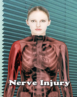
Brachial Plexus Injury
A network of nerves that conveys signals from the spine to arms, hands and shoulders is known as brachial plexus. Brachial refers to an arm and a network of nerves is indicated as plexus. Damage to these nerves causes brachial plexus injuries. A paralyzed arm, lack of muscle control in the arm, wrist or hand are some of the symptoms of brachial plexus injuries. Along with any of these symptoms, there is lack of feeling or sensation in the arm or hand.
Brachial plexus injuries commonly occur during birth but a possibility of this injury occurring at any time cannot be ruled out. When this injury happens during birth, the brachial plexus nerves stretch or tear as a result of an impact of the shoulders of the baby during the birth process. This injury is also known as Erb's Palsy when newborn is affected by it. A more serious condition called global palsy may occur when the injury involves both the upper and lower nerves. A difficult delivery when the baby is very large or if there is a breech or a protracted labor, pose the problem of brachial plexus injury. Brachial Plexus injuries are classified into four types:
- The most severe type is called avulsion. In this type of the injury, the nerve gets torn from the spine.
- The other type is called rupture. The nerve is torn in this type, but it occurs at a place other than the spinal attachment.
- The third type is called neuroma; in this type, a scar tissue will grow around the injury when the nerve tries to heal by itself and this exerts pressure on the injured nerve. It will also prevent the nerve from passing on signals to the muscles.
- The fourth type is neuropraxia or stretch. Though the nerve gets damaged due to the injury in this type it is not torn. Neuropraxia is the type of brachial plexus injury that occurs very commonly.
Occupational or physical therapy is available for treating brachial plexus injuries and some cases may need surgery. Recovery is not possible for avulsion and rupture injuries unless surgery is done well in time to reconnect the nerves. The potential for recovery differs from patient to patient, if the injury is either neuroma or neuropraxia. Recovery is spontaneous for patients who suffer from neuropraxia.
A new born affected with this injury will not be able to move the arm and will keep its arm stiff at its side. A more severe injury in an infant is indicated by a droopy eyelid on the affected side. The doctor will look for any damage to the bones and joints of the shoulder and the neck by ordering a x-ray or MRI. EMG is conducted to determine the presence of nerve signals in the muscles of the upper arm.
Since recovery is possible without surgery, most infants affected by Erb's palsy or brachial plexus injury will be examined after a month again. This is to confirm the extent of recovery of the nerves. The doctor will repeat this examination after two more months. For complete recovery, it may take even up to two years. The doctor may suggest a range of motion exercises that are most important to keep the infant's joints from getting stiff.
Neurotmesis
Neurotmesis etymology: Neurotmesis refers to most serious and severe nerve injury. Neurotmesis is brachial plexus injury. These brachial plexus injuries can occur in live births. The type of injury to the brachial plexus and the stretch damage will determine where the injury takes place. Various types of injuries can occur once the nerve rootlets form mixed nerve root. In some instances, the extent of the nerve damage may not be fully apparent but complete loss of motor, sensory and autonomic functions occurs. This type of complete rupture of the brachial plexus is called Neurotmesis. Neurotmesis is part of Seddon's classification scheme used to classify nerve damage. Seddon classified the nerve injury based on the extent of damage to the nerves on the basis of structural changes in cut nerves. The Seddon classification divides nerve injuries into three types namely:
Neurotmesis: Complete anatomic division of the nerve fibers with obvious discontinuity of the nerve sheath.

Axonotmesis: Microscopic division of nerve fibers without obvious discontinuity of nerve sheath.
Neuropraxia: There is injury without any anatomical discontinuity but resulting in functional disruption or nerve concussion. This is short term or sometimes lasts months with severe compression.
Neuropraxia Symptoms : Nerve Damage Symptoms: Common symptoms of Neurotmesis include loss of sensation and change in taste, expression and speech. There might be emotional and psychological disturbances. In the final stages, there could be a complete loss of motor, sensory and autonomic functions.
Diagnosis of Nerve Injury: There are many ways to diagnose the extent of the nerve injury. One of the common ways is Nerve conduction Velocity Test which tests the speed and strength of a signal being transmitted by nerve cells. Testing these factors can reveal the nature of nerve injury, such as damage to nerve cells or to the protective myelin sheath (protective coating on axons).
The test Electroneurography (EneG) which is also known as nerve conduction study or usually as a Nerve Conduction Velocity test(NCV) will help determine the nerve damage and further explore the choice of treatment.
Other than Peripheral nerve injuries, NCV is also helpful for the diagnosis of the following conditions:
Guillain Barré syndrome
Herniated disc disease
Charcot Marie Tooth disease
Special tests for assessment of Neurotmesis include electromyography, Strength duration curve, nerve conduction study and thermography. EMG test will be able to determine the presence, location and access the extent of diseases that caused the damage to the nerves and muscles. In some cases, a nerve biopsy may be needed where a small minute portion of the damaged nerve is surgically removed and analyzed.
Prognosis: Recovery from trauma is dependent on the age of the patient, type of injury and degree of injury. Without surgical intervention and repair this injury has very poor prognosis. Even with surgical repair, there could be significant loss of motor and sensory neurons which are responsible for normal conduction.
Tags: #Brachial Plexus Injury #Neurotmesis
At TargetWoman, every page you read is crafted by a team of highly qualified experts — not generated by artificial intelligence. We believe in thoughtful, human-written content backed by research, insight, and empathy. Our use of AI is limited to semantic understanding, helping us better connect ideas, organize knowledge, and enhance user experience — never to replace the human voice that defines our work. Our Natural Language Navigational engine knows that words form only the outer superficial layer. The real meaning of the words are deduced from the collection of words, their proximity to each other and the context.
Diseases, Symptoms, Tests and Treatment arranged in alphabetical order:

A B C D E F G H I J K L M N O P Q R S T U V W X Y Z
Bibliography / Reference
Collection of Pages - Last revised Date: February 16, 2026


